Basics

Principle of transmission electron tomography

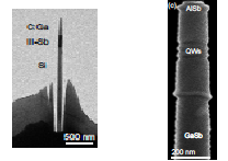
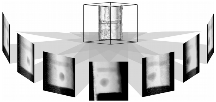
A needle-shaped sample cut out of any bulk material by a focused ion beam can be imaged in the microscope by sample tilting in several different orientations, resulting in a series of single projection images. In a second step, each tilted single-projection image can be back-projected in the computer in either real or reciprocal space to obtain a three-dimensional reconstruction of the original object.

For compact semiconductor structures, the sample is ideally needle-shaped along the tilt axis to best meet the projection requirements of electron tomography. The imaging mode is adapted to the problem, for example the HAADF-STEM (high-angle annular dark-field scanning TEM) method serves a chemically sensitive imaging and reveals compositional variations.
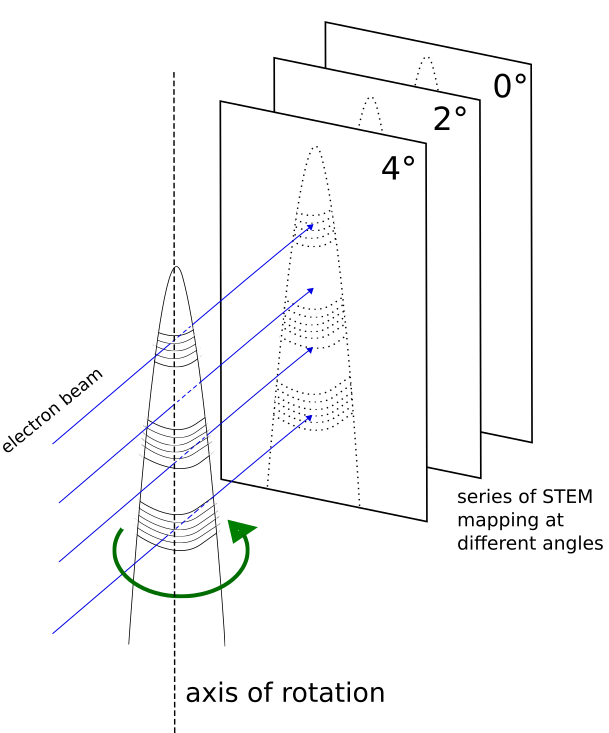
formation of projection
Tomogrammnadel
Tilt series
Reconstruction of a 3D object from the images of the tilt series.

Analysis and interpretation

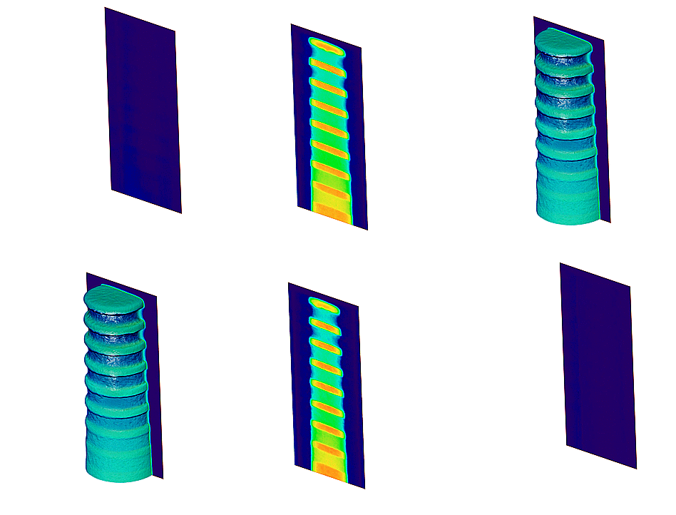
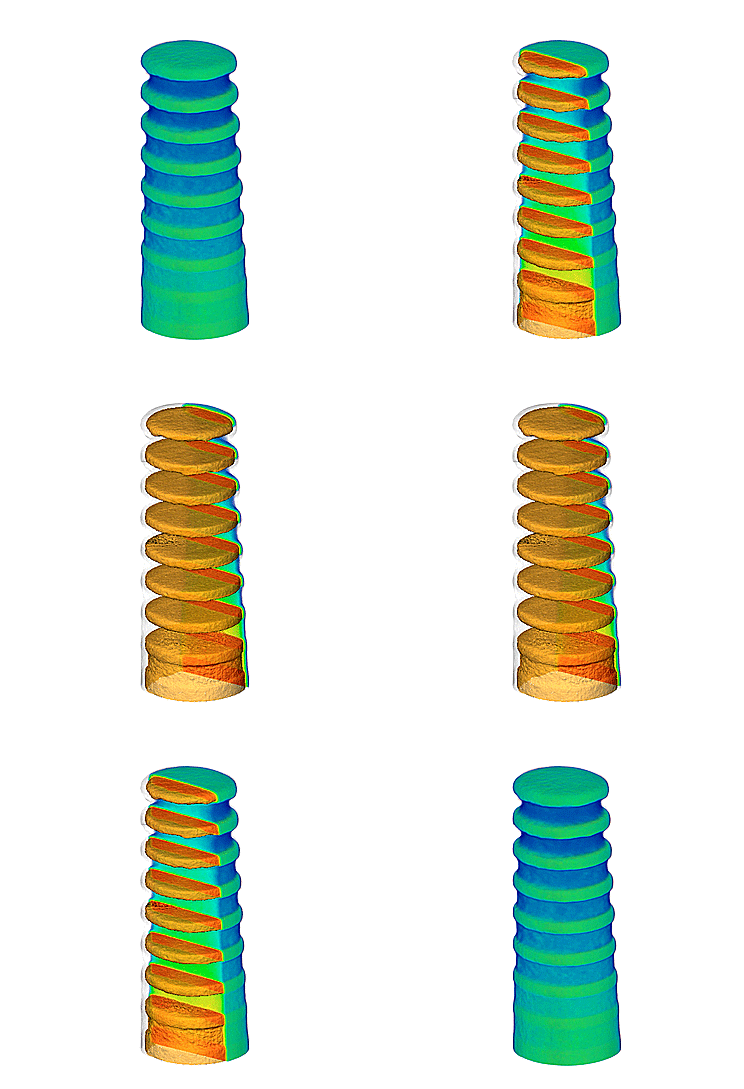
With modern software it is possible to visualize the reconstruction data in many ways and to display it three-dimensionally: the tomogram can be viewed from all spatial directions and by selective sections details inside can be made visible or highlighted for further analysis.

Example: Reconstruction of an epitaxial AlGaAs-GaAs interface to determine both its morphological roughness and the chemical width.

Funding

The "Application Laboratory Electron Tomography" is funded by the European Regional Development Fund (ERDF) of the European Union and by the State of Berlin (Project No. 2016011843).


Contact

Dr. Achim Trampert Foto
  • Dr. Achim Trampert
  • trampert@pdi-berlin.de
  • +49 (0)30 20377-280
  • Room 615
  • Hausvogteiplatz 5 - 7
    10117 Berlin
    Germany
  • Head of project